


❋
October 3-7 | Starting at $500Clinical Imaging Protocols & Order Templates
Staffing Request: Female technologist only; no students or observers.
Preparation: Fasting is required for abdominal and visceral vascular evaluations.
Important Note for All Studies:
1. Median Arcuate Ligament Syndrome (MALS) & SMA Stenosis
Order TitleWe offer a range of solutions designed to meet your needs—whether you're just getting started or scaling something bigger. Everything is tailored to help you move forward with clarity and confidence.Complete mesenteric arterial duplex ultrasound — fasting — median arcuate ligament syndrome dynamic respiratory protocol and superior mesenteric artery stenosis evaluation
Procedure CodeGetting started is simple. Reach out through our contact form or schedule a call—we’ll walk you through the next steps and answer any questions along the way.CPT 93975 — Complete visceral vascular duplex ultrasound
Diagnostic CodesICD-10-CM I77.4 — Celiac artery compression syndrome
ICD-10-CM K55.1 — Chronic vascular disorders of intestine / superior mesenteric artery stenosis
Protocol / Order Comments:
-
Evaluate the celiac artery for median arcuate ligament compression and the superior mesenteric artery (SMA) for stenosis.
-
Obtain angle-corrected peak systolic and end-diastolic velocities at:
Abdominal aorta
Celiac artery origin and proximal celiac artery
SMA origin, proximal, mid, and distal segments
-
Record celiac artery velocities during deep inspiration and complete end-expiration (both supine and upright).
-
Document respiratory excursion of the celiac artery, focal narrowing, turbulence, deflection/hooking, and post-stenotic dilatation.
-
Document SMA focal narrowing, elevated velocities, turbulence, post-stenotic changes, and velocity ratios relative to the abdominal aorta (when technically possible).
-
Collaborative, honest, and straightforward. We're here to guide the process, bring ideas to the table, and keep things moving.
-
Do not substitute a routine abdominal ultrasound.
2. Superior Mesenteric Artery (SMA) Syndrome
Order TitleLimited abdominal ultrasound — fasting — dynamic superior mesenteric artery syndrome protocolWe offer a range of solutions designed to meet your needs—whether you're just getting started or scaling something bigger. Everything is tailored to help you move forward with clarity and confidence.Complete mesenteric arterial duplex ultrasound — fasting — median arcuate ligament syndrome dynamic respiratory protocol and superior mesenteric artery stenosis evaluation
Procedure CodeCPT 76705 — Limited abdominal ultrasoundGetting started is simple. Reach out through our contact form or schedule a call—we’ll walk you through the next steps and answer any questions along the way.CPT 93975 — Complete visceral vascular duplex ultrasound
Diagnostic CodesICD-10-CM K55.1 — Superior mesenteric artery syndrome
Protocol / Order Comments:
-
Evaluate the celiac artery for median arcuate ligament compression and the superior mesenteric artery (SMA) for stenosis.
-
In the sagittal plane, measure and document:
Aortomesenteric angle
Shortest aortomesenteric distance
-
Assess the third portion of the duodenum for focal compression. Evaluate for proximal duodenal/gastric dilation and impaired passage through the compressed segment.
-
Dynamically assess duodenal passage and compression changes across multiple positions: supine, upright, left-lateral, and prone (when technically feasible).
-
Use color Doppler as needed to identify the abdominal aorta and SMA.
-
Collaborative, honest, and straightforward. We're here to guide the process, bring ideas to the table, and keep things moving.
-
This is an anatomic evaluation for duodenal compression and is separate from the mesenteric arterial duplex evaluation for SMA stenosis.
3. Nutcracker Syndrome
Order TitleComplete renal venous duplex ultrasound — fasting — Nutcracker syndrome
Procedure CodeCPT 93975 — Complete visceral vascular duplex ultrasound
Diagnostic CodesICD-10-CM I87.1 — Compression of vein
Protocol / Order Comments:
-
Evaluate the left renal vein (LRV) from the renal hilum through the aortomesenteric segment to its junction with the inferior vena cava (IVC).
-
In the sagittal plane, measure and document the aortomesenteric angle and shortest aortomesenteric distance. Correlate with the location and severity of LRV compression.
Measure LRV diameter and peak velocity at: the renal hilum, immediately proximal to compression, within the aortomesenteric compression, and distal to compression.
Calculate hilar-to-aortomesenteric diameter and velocity ratios.
-
Perform imaging in both supine and upright positions if technically feasible.
-
Assess for anterior, posterior, retroaortic, or circumaortic LRV compression anatomy; reversed/abnormal flow; collateral veins; and left ovarian-vein dilatation or reflux.
4. Inferior Vena Cava (IVC) Compression
Order TitleComplete inferior vena cava venous duplex ultrasound — fasting — dynamic positional and respiratory compression protocolComplete renal venous duplex ultrasound — fasting — Nutcracker syndrome
Procedure CodeCPT 93978 — Complete duplex ultrasound of the aorta, inferior vena cava, and/or iliac vasculatureCPT 93975 — Complete visceral vascular duplex ultrasound
Diagnostic CodesICD-10-CM I87.1 — Compression of vein / acquired vena cava stenosisICD-10-CM I87.1 — Compression of vein
Protocol / Order Comments:
-
Evaluate the entire visualized IVC for patency, thrombus, focal stenosis, extrinsic compression, obstruction, and collateralization.
-
Perform imaging supine and upright (use seated or reverse-Trendelenburg if upright is limited).
Include Valsalva maneuvers where technically appropriate.
-
Record vessel diameters, flow direction, Doppler waveforms, and velocities at the proximal, mid, and distal IVC during quiet respiration, deep inspiration, and complete end-expiration.
-
Document focal narrowing, pre- and post-compression velocities, velocity ratios, continuous or nonphasic flow, collateral veins, and any non-visualized segments.
5. Bilateral Iliac-Vein Compression / Nonthrombotic Iliac-Vein Lesions
Order TitleComplete bilateral iliac venous duplex ultrasound — fasting — bilateral iliac-vein compression and nonthrombotic iliac-vein lesion protocol
Procedure CodeCPT 93978 — Complete duplex ultrasound of the aorta, inferior vena cava, and/or iliac vasculature
Diagnostic CodesICD-10-CM I87.1 — Compression of vein / iliac-vein compression syndromeICD-10-CM I87.1 — Compression of vein / iliac-vein compression syndrome
Protocol / Order Comments:
-
Evaluate right and left common iliac veins and external iliac veins for nonthrombotic iliac-vein lesions (NIVLs), focal stenosis, extrinsic arterial compression, obstruction, thrombus, and collateralization.
-
Include the IVC confluence and bilateral common femoral veins.
-
Specifically evaluate for classic left common iliac-vein compression by the right common iliac artery (May-Thurner anatomy) as well as right-sided, bilateral, or variant compression.
-
Obtain diameter measurements and Doppler velocities proximal to, at, and distal to each suspected compression. Calculate diameter-reduction and velocity ratios.
-
Perform supine imaging and repeat in upright, seated, or reverse-Trendelenburg positioning when feasible. Document any non-visualized segments.
6. Chronic Venous Insufficiency (CVI)5. Bilateral Iliac-Vein Compression / Nonthrombotic Iliac-Vein Lesions
Order TitleBilateral lower-extremity complete venous reflux duplex ultrasoundComplete bilateral iliac venous duplex ultrasound — fasting — bilateral iliac-vein compression and nonthrombotic iliac-vein lesion protocol
Procedure CodeCPT 93970 — Complete bilateral extremity venous duplex ultrasound
Diagnostic CodesICD-10-CM I87.2 — Venous insufficiency, chronic, peripheral
Protocol / Order Comments:
-
Perform with the patient standing or in a dependent position.
-
Evaluate deep, superficial, and clinically relevant perforator veins for obstruction and reflux (including common femoral, femoral, deep femoral, popliteal, great saphenous, small saphenous, and accessory saphenous veins).
-
Use Valsalva and distal augmentation. Document the location and duration of reflux and provide bilateral vein mapping.
-
Obtain diameter measurements and Doppler velocities proximal to, at, and distal to each suspected compression. Calculate diameter-reduction and velocity ratios.
-
This is a complete venous reflux study, not a deep-vein-thrombosis-only (DVT) clot check.

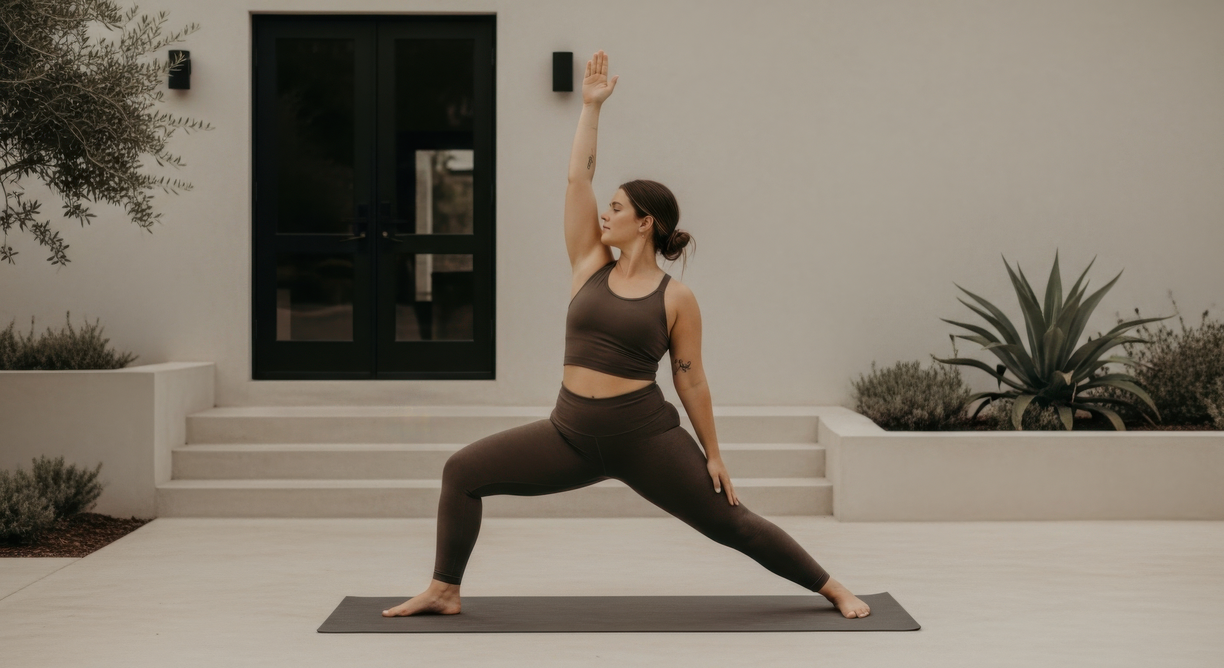
Reconnect with your body and mind as you escape the noise of everyday life.
Day OneArrive & Meet
As everyone arrives, we take time to settle in, get comfortable, and begin connecting with those around us. The journey starts here.
Check-In9:00 – 9:30am
Group Activity11:00am
Lunch Break12:30pm
Creative Workshop2:00pm
Dinner6:30pm

Day TwoSet Intentions & Reflect
Together, we pause to consider our goals, hopes, and direction. This is about aligning with ourselves and with the journey ahead. This is a chance to reconnect with what brought you here—your questions, your hopes, your turning points—and consider how they’ve shifted or deepened.
Check-In9:00 – 9:30am
Group Activity11:00am
Lunch Break12:30pm
Creative Workshop2:00pm
Dinner6:30pm

Day ThreeLook Forward & Wrap Up
We explore the possibilities beyond this moment, making space for growth, action, and forward momentum. As we end our time together, we honor the experience, the growth, and the connections made along the way.
Check-In9:00 – 9:30am
Group Activity11:00am
Lunch Break12:30pm
Creative Workshop2:00pm
Dinner6:30pm
How It Works-
Browse our upcoming events to find the one that feels right for you. We host events year-round in all different locations and climates.
-
Sign up and pay all required fees to reserve your spot. If plans change, you can cancel up to 14 days before the retreat start to receive a 50% refund.
-
After booking, we'll send you a Welcome Packet with everything you need to know—detailed schedules, packing list recommendations, add-ons to consider, and more.
-
We'd recommend booking your transportation to and from the event as soon as possible, to ensure you can arrive without any complications or delays.
-
Now all that's left to do is pack your bags and get excited for your new adventure.